W.M. Keck Foundation
Scanning Electron Microscope
The W.M. Keck Electron Microscopy Center utilizes the SEM in a number of different research projects and courses.

Deanna Soper, Ph.D.
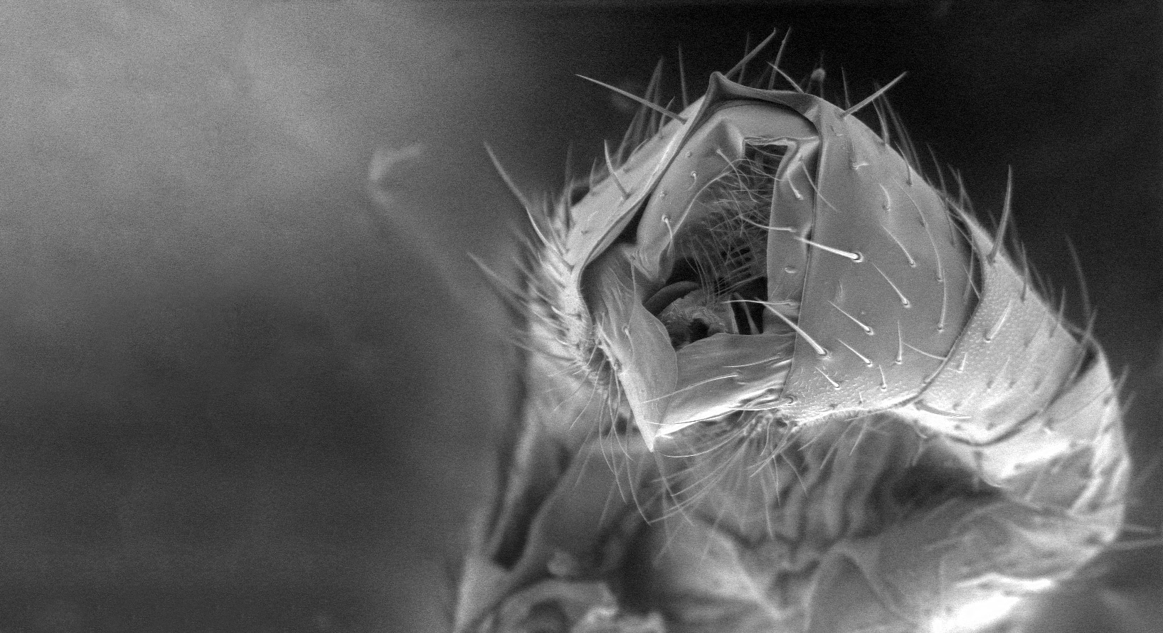
The Soper Lab is using the SEM microscope to better understand both morphological structures and physiological processes. We use the SEM to examine the genitalia of fruit flies and snails. Genitalia is frequently analyzed by biologists because it is a quickly evolving trait, which can lead to speciation. Frequently, especially in insects, it is the only trait taxonomists can use to distinguish different species. In addition, we have a collaboration with Mote Marine Laboratory to examine the effects of increased ocean temperature and decreased pH on stony coral skeleton formation. The SEM has the capability to quantify elemental content, and we are using that data to determine if coral skeleton formation is altered under these two different environmental parameters.
Learn more about Dr. Soper
Ellen Steinmiller, Ph.D.
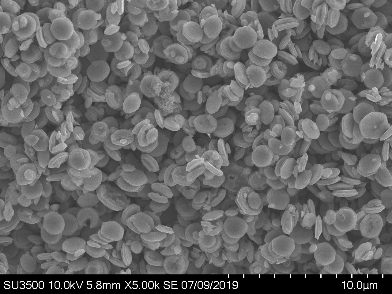
The SEM is used in the W.M. Keck Electron Microscopy Center to investigate the morphology of composite inorganic photocatalysts. The SEM allows both imaging and elemental analysis of the synthesized materials. The W.M. Keck Electron Microscopy Center also uses the SEM to image bacteria in a collaboration with the Cody Lab in the Biology Department.
Learn more about Dr. Steinmiller
, Laboratory Manager Arthur Sweeney (Physics), and Associate Professor Ellen Steinmiller (Chemistry) are working to demo the scope for prospective students.")
Assistant Professor Deanna Soper (Biology), Laboratory Manager Arthur Sweeney (Physics), and Associate Professor Ellen Steinmiller (Chemistry) are working to demo the scope for prospective students.
 attachment that allows researchers to quantify elemental composition.")
Energy Dispersive X-Ray Analyzer (EDX) attachment that allows researchers to quantify elemental composition.

Dr. Steinmiller showing Arthur Sweeney and a UD student elemental analysis using the EDX.

ZnO "rods" synthesized in the W.M. Keck Electron Microscopy Center.

Drosophila melanogaster male genitalia. Photo credit: Dr. Deanna Soper

Pseudomonas, a common bacteria found all over the world in soil, water and plants.
. Photo credit: Dr. Deanna Soper")
Potamopyrgus antipodarum, a freshwater snail from New Zealand head and genitalia (on top).
Photo credit: Dr. Deanna Soper
Contact Us
dsoper@udallas.edu | 972-721-5245
esteinmiller@udallas.edu | 972-721-5110